Physical Therapy for Patellofemoral Pain: Expert Tips
Patellofemoral pain syndrome (PFPS) affects millions of individuals worldwide, causing discomfort around the kneecap and limiting daily activities. This condition, commonly experienced by athletes, office workers, and active individuals, occurs when the patella (kneecap) does not track properly along the femur (thighbone), leading to pain and dysfunction. Understanding the mechanisms behind patellofemoral syndrome and implementing evidence-based physical therapy interventions can significantly reduce symptoms and restore functional mobility.
The journey to recovery from patellofemoral pain requires a comprehensive approach combining therapeutic exercises, manual techniques, and lifestyle modifications. Physical therapists employ specialized assessment methods and targeted interventions to address the underlying biomechanical dysfunctions contributing to knee pain. This guide explores expert-recommended strategies for managing patellofemoral syndrome effectively.
Understanding Patellofemoral Pain Syndrome
Patellofemoral pain syndrome represents a multifactorial condition involving complex interactions between muscular weakness, biomechanical dysfunction, and movement patterns. The condition develops when cumulative stress on the patellofemoral joint exceeds tissue tolerance, resulting in inflammation and pain around the anterior knee. Research indicates that weakness in hip abductors and external rotators significantly contributes to altered knee mechanics and increased patellofemoral stress.
The etiology of PFPS typically involves several interconnected factors including decreased hip strength, poor core stability, excessive foot pronation, and inadequate vastus medialis obliquus (VMO) activation. Unlike structural knee injuries, patellofemoral pain often results from functional deficits that respond exceptionally well to targeted physical therapy. Understanding these underlying mechanisms enables therapists to design individualized treatment programs addressing root causes rather than merely symptom management.
Clinical evidence demonstrates that comprehensive therapy resources and information emphasizing biomechanical correction yield superior long-term outcomes compared to isolated pain management approaches. The condition affects various populations differently, requiring personalized assessment and intervention strategies.

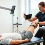
Assessment and Diagnosis Methods
Accurate assessment forms the foundation of effective patellofemoral syndrome physical therapy. Physical therapists utilize multiple evaluation techniques including patient history analysis, functional movement screening, and specialized orthopedic tests to identify contributing factors. The comprehensive assessment process examines hip strength, knee alignment, ankle mobility, and lower limb kinetic chain function.
Key assessment components include:
- Hip Strength Testing: Evaluating abductor and external rotator strength using manual muscle testing and dynamometry to identify significant deficits contributing to knee pain
- Knee Alignment Analysis: Assessing Q-angle, tibial rotation, and patellar tracking during dynamic movements using functional tests like single-leg squats
- Ankle and Foot Evaluation: Examining dorsiflexion mobility, pronation patterns, and arch support to determine lower chain contributions to pain
- Movement Quality Screening: Observing movement patterns during stairs, lunges, and running to identify compensatory mechanisms
- Strength and Flexibility Assessment: Measuring quadriceps, hamstring, and calf flexibility alongside hip and core strength levels
Advanced diagnostic imaging including MRI or ultrasound may supplement clinical assessment when structural pathology is suspected. However, research emphasizes that clinical assessment findings correlate more strongly with functional outcomes than imaging results alone. Evidence-based clinical guidelines recommend prioritizing functional assessment over imaging for treatment planning.
Core Strengthening Exercises
Core stability forms the foundation for proper lower limb mechanics and pain-free movement. The core encompasses abdominal, lumbar, and hip musculature working synergistically to stabilize the spine and pelvis during dynamic activities. Weakness in these structures forces compensatory patterns that increase patellofemoral stress and perpetuate pain cycles.
Essential core exercises for patellofemoral pain management include:
- Plank Variations: Standard planks, side planks, and quadruped holds build isometric core endurance necessary for stability during weight-bearing activities
- Glute Bridges: Activating gluteal muscles and teaching hip extension patterns critical for proper knee mechanics during walking and running
- Dead Bugs: Coordinating core activation with limb movement to develop neuromuscular control patterns transferable to functional activities
- Pallof Presses: Resisting rotational forces to strengthen lateral core stabilizers and improve anti-rotation stability
- Bird Dogs: Developing contralateral limb coordination and core endurance through alternating arm and leg extension
Progressive core training incorporates increasing difficulty through additional resistance, unstable surfaces, and integrated movement patterns. Therapists emphasize quality execution over quantity, ensuring proper muscle activation patterns develop. The relationship between core stability and knee health demonstrates why comprehensive core training precedes advanced strengthening protocols.
Lower Limb Alignment Correction
Correcting lower limb alignment deficits represents a critical component of patellofemoral syndrome rehabilitation. Hip weakness causes the knee to collapse inward during weight-bearing activities (dynamic knee valgus), increasing patellofemoral compression forces. Similarly, excessive foot pronation alters knee mechanics and contributes to pain development.
Hip strengthening protocols specifically target abductors and external rotators through progressive resistance exercises:
- Clamshells: Isolating gluteus medius activation and external rotation strength in side-lying positions
- Side-Lying Hip Abduction: Building hip abductor strength essential for controlling knee position during single-leg activities
- Monster Walks: Combining hip abduction with forward progression to develop functional strength patterns
- Lateral Band Walks: Using elastic resistance to strengthen hip abductors during functional movement patterns
- Single-Leg Stance: Developing dynamic hip stability necessary for maintaining proper knee alignment
Foot and ankle mobility corrections address excessive pronation through calf stretching, intrinsic foot strengthening, and potentially custom orthotics. The kinetic chain relationship means that ankle restriction forces compensatory knee mechanics. Therefore, physical therapy treatment approaches increasingly recognize the importance of comprehensive lower limb assessment and intervention.
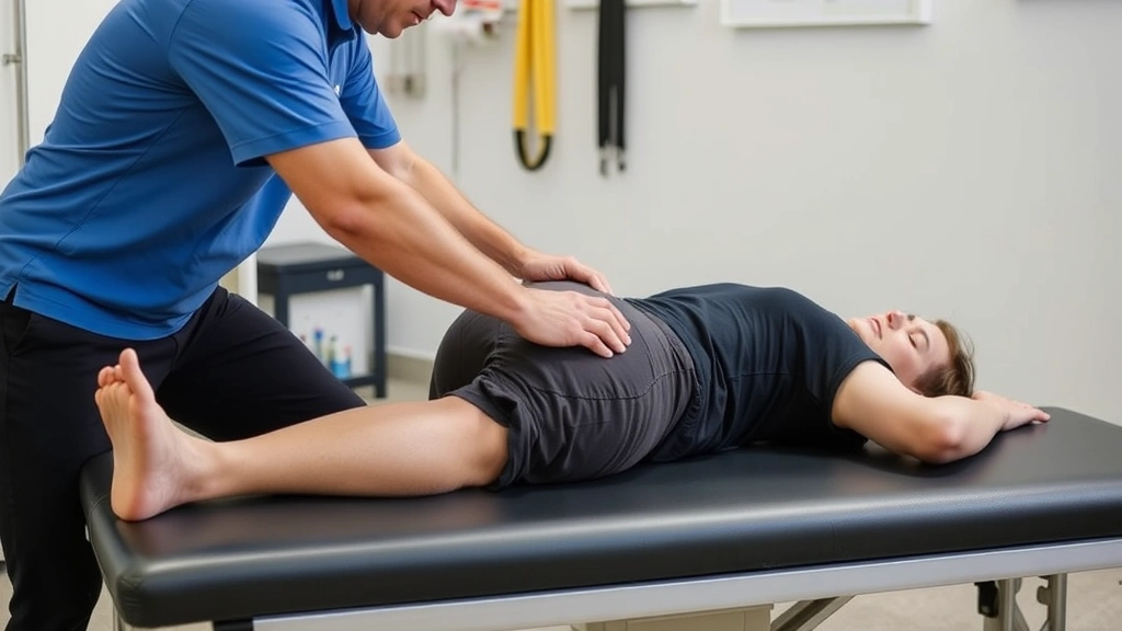
Soft Tissue Mobilization Techniques
Soft tissue restrictions in muscles, fascia, and connective tissues contribute to altered movement mechanics and patellofemoral pain. Physical therapists employ manual therapy techniques combined with self-mobilization strategies to restore tissue extensibility and movement quality.
Key soft tissue mobilization approaches include:
- Myofascial Release: Using sustained pressure on muscle trigger points and fascial restrictions to reduce tension and improve tissue quality
- Instrument-Assisted Soft Tissue Mobilization (IASTM): Employing specialized instruments to address scar tissue and chronic restrictions in muscles surrounding the knee
- Foam Rolling: Self-directed myofascial release targeting quadriceps, iliotibial band, and hip muscles to reduce tension
- Manual Stretching: Therapist-directed stretching of tight structures including hip flexors, quadriceps, and hamstrings
- Joint Mobilization: Therapeutic movements of the patellofemoral and tibiofemoral joints to improve arthrokinematics and reduce pain
Research demonstrates that combining soft tissue mobilization with strengthening exercises produces superior outcomes compared to isolated approaches. The tissue extensibility improvements achieved through mobilization enable patients to execute exercises with proper form and full range of motion, accelerating recovery timelines.
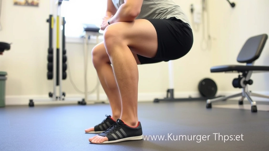
Progressive Loading Protocols
Successful patellofemoral syndrome management requires systematic progression from pain-free basic exercises toward challenging functional activities. Loading protocols must carefully balance stimulus provision with tissue tolerance to promote adaptation without exacerbating symptoms. This evidence-based approach prevents compensatory patterns that perpetuate pain cycles.
Progressive loading follows a structured framework:
Phase 1 (Weeks 1-2): Pain-free range strengthening focusing on isometric holds, controlled movements, and basic stability exercises. Patients establish baseline strength and learn proper movement patterns without provocative loading.
Phase 2 (Weeks 3-4): Introducing light resistance through bodyweight exercises and elastic bands. Progressively increasing repetitions and sets while maintaining movement quality. Patients begin single-leg activities and weight-shifting exercises.
Phase 3 (Weeks 5-6): Adding significant resistance through weights, increased repetitions, and more challenging positions. Introducing dynamic balance activities and sport-specific movement patterns. Patients perform exercises requiring greater neuromuscular control.
Phase 4 (Weeks 7-8+): Sport-specific training, plyometric activities, and high-demand functional tasks. Integrating exercise into sport or activity contexts with progressive intensity and complexity.
This systematic progression approach allows tissue adaptation while preventing overload. Monitoring pain patterns and movement quality guides progression decisions, ensuring patients advance appropriately. The relationship between therapy modalities and types emphasizes how progressive loading represents a fundamental principle across effective rehabilitation programs.
Return to Activity Guidelines
Returning to previous activity levels represents the ultimate goal of patellofemoral syndrome rehabilitation. However, premature return to high-demand activities frequently results in symptom recurrence and setback. Evidence-based return-to-activity protocols establish clear criteria and progressive milestones ensuring successful long-term recovery.
Return-to-activity criteria typically include:
- Pain-Free Strength: Achieving hip and core strength measurements within 90% of the unaffected side using objective testing
- Movement Quality: Demonstrating proper mechanics during functional movements without compensation patterns or dynamic knee valgus
- Functional Performance: Completing single-leg hop tests, lateral bounds, and sport-specific movements with confidence and proper form
- Symptom Resolution: Maintaining pain-free status during progressively demanding activities over extended timeframes
- Psychological Readiness: Developing confidence in knee stability and pain-free movement capacity
Sport or activity-specific training bridges the gap between rehabilitation exercises and actual performance demands. Athletes gradually reintroduce sport-specific movements, positions, and intensities while monitoring symptoms. Running progression typically begins with walk-run intervals progressing toward continuous running at varied paces and distances.
Long-term success requires maintaining strength and movement quality through ongoing exercise participation. Research indicates that continued exercise participation significantly reduces symptom recurrence and supports durable outcomes. Many individuals benefit from ongoing maintenance programs emphasizing hip and core strengthening.
Frequently Asked Questions
How long does patellofemoral pain recovery typically require?
Recovery timelines vary considerably based on pain severity, patient compliance, and individual factors. Most individuals experience significant improvement within 4-6 weeks of consistent physical therapy. However, complete resolution often requires 8-12 weeks or longer, particularly for chronic cases. Some individuals benefit from extended treatment addressing persistent deficits. Regular exercise participation following formal therapy completion prevents symptom recurrence.
Can I continue exercising with patellofemoral pain?
Yes, modified exercise participation accelerates recovery compared to complete rest. The key involves identifying pain-free activities and gradually building tolerance through progressive loading. Walking, swimming, and cycling often remain tolerable while running and jumping may require temporary modification. Physical therapists guide activity selection ensuring exercises promote recovery rather than exacerbate symptoms.
Do I need custom orthotics for patellofemoral pain?
Custom orthotics benefit some patients with significant foot pronation or structural foot issues contributing to pain. However, research suggests that many individuals achieve full recovery through exercise alone without orthotics. A physical therapist can assess whether orthotics may benefit your specific situation. Off-the-shelf arch supports sometimes provide adequate support at lower cost.
What exercises should I avoid with patellofemoral pain?
Exercises causing pain should be temporarily avoided or modified. Common provocative activities include deep squats, lunges, stairs, and running. However, modified versions of these movements often remain tolerable. Pain-free strengthening exercises form the foundation while gradually building tolerance to previously painful activities through progressive loading.
Is patellofemoral pain permanent?
No, patellofemoral pain is not permanent and responds exceptionally well to appropriate physical therapy. The majority of individuals achieve complete symptom resolution through consistent exercise and biomechanical correction. However, ongoing maintenance exercise participation prevents recurrence. Understanding that recovery requires active participation and sustained effort optimizes long-term outcomes.
How does patellofemoral pain differ from other knee injuries?
Patellofemoral pain typically results from functional deficits and biomechanical dysfunction rather than structural damage to cartilage or ligaments. This distinction means PFPS responds particularly well to physical therapy and exercise-based rehabilitation. Imaging often appears normal despite significant symptoms. The functional nature of the condition makes it highly treatable through targeted intervention addressing underlying causes.
Should I see a physical therapist for patellofemoral pain?
Yes, physical therapy represents the gold standard treatment for patellofemoral pain syndrome. A qualified physical therapist conducts comprehensive assessment identifying contributing factors and designs individualized treatment programs. Early intervention typically produces faster recovery and prevents chronic pain development. Many insurance plans cover physical therapy with appropriate physician referral, and understanding therapy costs helps individuals plan treatment access.
Can I prevent patellofemoral pain recurrence?
Yes, maintaining hip and core strength through ongoing exercise participation significantly reduces recurrence risk. Gradual progression when increasing activity intensity or volume prevents overload. Attention to movement quality during daily activities and sports maintains proper biomechanics. Regular stretching and mobility work preserve tissue extensibility. Many individuals benefit from periodic physical therapy sessions addressing emerging deficits before symptoms develop.