Physical Therapy for Patellofemoral Syndrome: Expert Insights & Evidence-Based Treatment
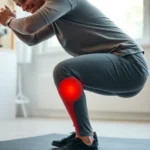
Patellofemoral syndrome (PFS), commonly known as runner’s knee, affects millions of individuals across all activity levels. This condition involves pain around the kneecap (patella) and is particularly prevalent among athletes, active individuals, and those with sedentary lifestyles. The good news is that physical therapy for patellofemoral syndrome has demonstrated remarkable effectiveness in managing symptoms and restoring function without requiring surgical intervention in most cases.
Understanding the biomechanical factors underlying patellofemoral syndrome is crucial for effective treatment. Unlike acute injuries, PFS develops gradually through repetitive stress, muscle imbalances, and improper movement patterns. This comprehensive guide explores expert-backed physical therapy approaches, evidence-based interventions, and practical strategies to overcome patellofemoral syndrome and return to pain-free movement.
Understanding Patellofemoral Syndrome
Patellofemoral syndrome represents a complex condition characterized by anterior knee pain that worsens with specific activities. Unlike structural damage visible on imaging, PFS involves functional movement impairments and muscular imbalances. The condition typically manifests during activities that load the knee joint, including running, jumping, squatting, and stair climbing.
The prevalence of patellofemoral syndrome spans across demographics. Runners report the highest incidence rates, with studies indicating that 7-10% of the general population experiences PFS symptoms. Interestingly, the condition affects women at nearly twice the rate of men, largely due to biomechanical differences in hip and knee anatomy.
Pain patterns in PFS vary considerably. Some individuals experience sharp, localized pain directly beneath the kneecap, while others report diffuse discomfort around the patella. Pain typically intensifies with prolonged sitting (theater sign), ascending or descending stairs, and explosive movements. The chronic nature of untreated PFS can significantly impact quality of life and athletic performance, making timely intervention essential.
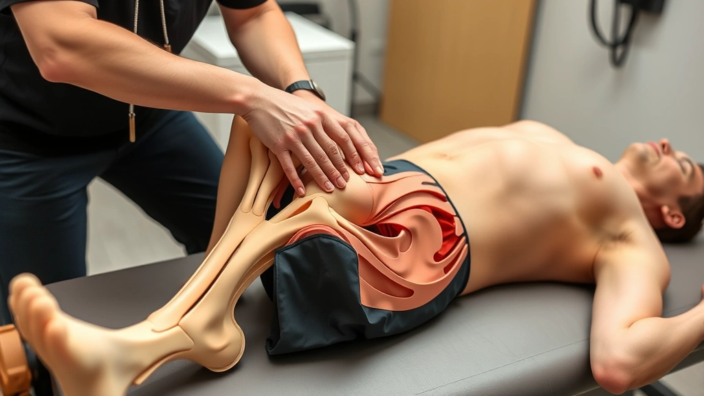
Biomechanical Factors and Root Causes
The biomechanics of patellofemoral syndrome involve complex interactions between hip stability, knee alignment, ankle mobility, and core function. Physical therapists recognize that treating the knee alone often proves insufficient—successful management requires addressing the entire lower kinetic chain.
Hip weakness and dysfunction represent primary contributors to PFS. The gluteus medius, gluteus maximus, and hip external rotators stabilize the pelvis and control femoral internal rotation during weight-bearing activities. When these muscles weaken, the femur internally rotates excessively, altering the patellar tracking mechanism and increasing stress on patellofemoral cartilage.
Quadriceps muscle imbalances also play significant roles. The vastus medialis obliquus (VMO) provides dynamic patellar stabilization, particularly during early knee flexion. Weakness or inhibition of the VMO relative to the vastus lateralis creates lateral patellar tracking, increasing compression forces on lateral facets.
Additional biomechanical factors include:
- Ankle mobility restrictions forcing compensatory knee valgus during squatting movements
- Excessive foot pronation causing internal tibial rotation and altered knee mechanics
- Core instability reducing proximal stability during dynamic activities
- Patellar tracking abnormalities from tight lateral structures and weak medial stabilizers
- Training errors including rapid increases in volume, intensity, or frequency
Understanding these multifactorial causes enables physical therapists to develop targeted interventions addressing specific impairments rather than applying generic treatment protocols.
If you’re exploring broader therapeutic approaches, our therapy resources provide additional insights into various rehabilitation methodologies.
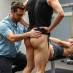
Physical Therapy Assessment and Diagnosis
Comprehensive assessment forms the foundation of effective patellofemoral syndrome physical therapy. Skilled physical therapists conduct thorough evaluations examining movement patterns, muscular strength and flexibility, biomechanical alignment, and functional limitations.
Initial evaluations typically include:
- Patient history and symptom analysis identifying pain triggers, onset patterns, and functional limitations
- Postural assessment evaluating spinal alignment, pelvic position, knee valgus, and foot pronation
- Range of motion testing measuring hip, knee, and ankle mobility
- Muscle strength testing using manual muscle testing and dynamometry to identify weakness patterns
- Movement screening observing squatting mechanics, step-down patterns, and gait analysis
- Special testing including patellar mobility assessment, McConnell test, and lower extremity alignment evaluation
- Functional testing replicating activities that reproduce symptoms
Research from the Journal of Orthopaedic & Sports Physical Therapy demonstrates that individualized assessment-driven interventions yield superior outcomes compared to standardized protocols. Physical therapists identify specific impairments driving each patient’s symptoms, enabling precision medicine approaches to treatment.
” alt=”Physical therapist performing knee assessment on patient in clinical setting”>
Core Treatment Strategies
Evidence-based physical therapy for patellofemoral syndrome emphasizes systematic impairment correction through progressive exercise programming. Treatment typically progresses through three distinct phases: pain management and protection, restoration of function, and return to activity.
Phase 1: Pain Management and Protection
Initial treatment focuses on reducing pain through activity modification and therapeutic modalities. Physical therapists recommend temporarily avoiding movements that reproduce symptoms while introducing pain-free exercises. This strategic approach maintains neuromuscular activation without exacerbating inflammation.
Therapeutic modalities may include:
- Ice application for acute inflammation management
- Electrical stimulation modalities reducing pain perception
- Manual therapy techniques addressing soft tissue restrictions
- Patellar taping or bracing optimizing biomechanics during healing
Phase 2: Impairment Correction
The primary treatment phase addresses specific biomechanical impairments identified during assessment. This typically involves progressive strengthening of hip abductors and external rotators, quadriceps (particularly VMO), and core stabilizers. Concurrent flexibility work addresses tight muscles contributing to altered mechanics.
Physical therapists utilize various training methods including:
- Isometric exercises developing strength without movement
- Isotonic exercises through controlled ranges of motion
- Isokinetic training optimizing strength across movement speeds
- Functional training integrating strength gains into movement patterns
For additional insights into specialized therapeutic approaches, explore our information on red light therapy and other complementary modalities that some practitioners incorporate into comprehensive treatment plans.
Phase 3: Return to Activity
Once strength and pain improve, progressive return to running, sports, or recreational activities follows carefully structured protocols. This phase emphasizes movement quality, gradual intensity progression, and appropriate recovery between sessions.
Strengthening Exercises for Patellofemoral Syndrome
Targeted strengthening represents the cornerstone of patellofemoral syndrome physical therapy. Research consistently demonstrates that progressive resistance exercise addressing hip abductors, hip external rotators, and quadriceps yields superior long-term outcomes.
Hip Abductor Strengthening
The gluteus medius functions as the primary hip abductor and internal rotator controller. Effective exercises include:
- Clamshells in side-lying position, progressing to standing hip abduction with resistance bands
- Side-lying leg lifts targeting hip abductors through gravity-resisted movement
- Monster walks with resistance bands around knees, emphasizing controlled movement quality
- Single-leg stance variations developing dynamic pelvic stability
- Lateral band walks maintaining tension throughout movement
Hip External Rotator Strengthening
External rotation strength prevents femoral internal rotation during weight-bearing activities:
- Clam exercises isolating external rotators in side-lying position
- Standing external rotation with resistance bands mimicking functional movement patterns
- Four-way hip exercises addressing all hip movement directions systematically
- Single-leg deadlifts integrating external rotation stability into functional movement
Quadriceps Strengthening
VMO-focused quadriceps strengthening optimizes patellar tracking:
- Short-arc quadriceps sets activating quadriceps without full knee extension stress
- Straight leg raises with hip external rotation emphasis
- Terminal knee extension exercises strengthening VMO through final extension range
- Wall sits with hip abduction combining quadriceps and hip abductor activation
- Step-ups and step-downs progressing to full functional weight-bearing exercises
Core and Trunk Stabilization
Proximal stability enables efficient lower extremity function:
- Planks and side planks developing core endurance
- Dead bugs and bird dogs coordinating trunk and extremity movement
- Bridging exercises activating gluteal muscles and core stabilizers
- Pallof presses addressing anti-rotation stability crucial for running mechanics
Physical therapists progress exercises systematically, increasing resistance, complexity, and functional relevance as tolerance improves. Research published by The American Physical Therapy Association emphasizes that exercise adherence and progressive overload prove more important than specific exercise selection for long-term success.
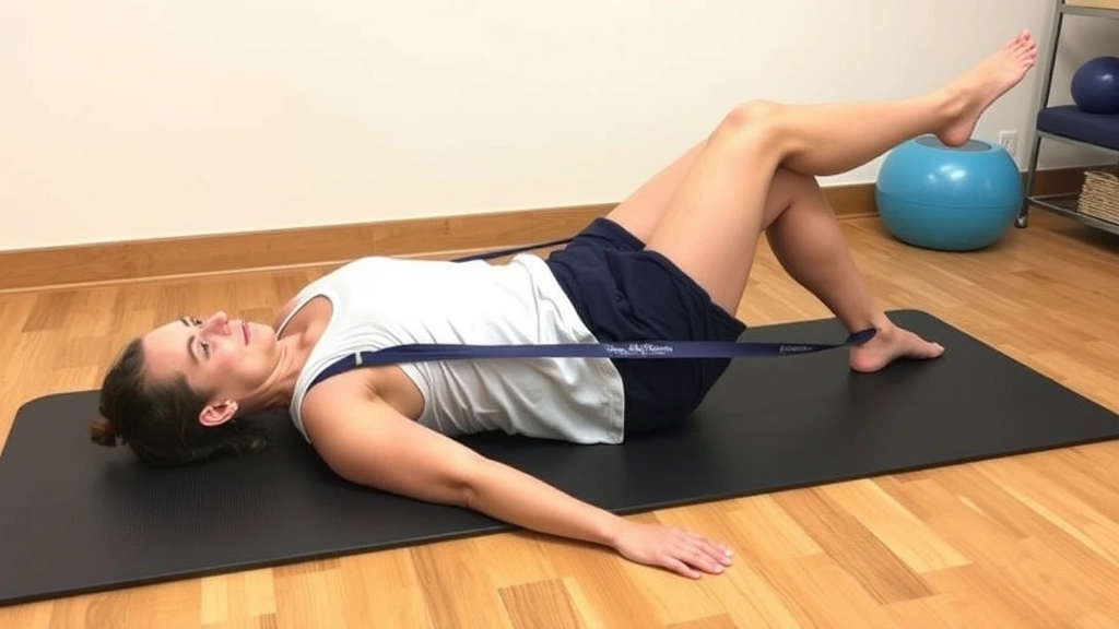
Flexibility and Mobility Work
While strengthening receives primary emphasis, flexibility and mobility work addresses muscle tightness contributing to altered biomechanics. Physical therapists identify specific structures requiring mobility work based on individual assessment findings.
Hip Flexor Stretching
Tight hip flexors (iliopsoas) reduce hip extension range of motion, causing compensatory lumbar extension and altered knee mechanics during running. Effective stretches include:
- Kneeling hip flexor stretches emphasizing anterior hip elongation
- Half-kneeling position variations allowing progressive stretch intensity
- Dynamic stretching through controlled hip extension movements
IT Band and Lateral Thigh Mobility
The iliotibial band often becomes tight in PFS, contributing to lateral knee tracking abnormalities:
- Foam rolling addressing myofascial restrictions
- Cross-body IT band stretches isolating lateral thigh structures
- Soft tissue mobilization techniques releasing adhesions
Ankle and Calf Mobility
Restricted ankle dorsiflexion forces compensatory knee valgus during squatting and running. Mobility work includes:
- Dorsiflexion stretching with resistance band assistance
- Ankle mobilization exercises improving joint mechanics
- Calf stretching addressing plantarflexor tightness

” alt=”Patient performing stretching and flexibility exercises with physical therapist guidance”>
Return to Activity Protocols
Returning to running, sports, or high-impact activities requires carefully structured progression preventing reinjury. Physical therapists implement evidence-based return-to-activity protocols emphasizing gradual load increases and movement quality.
Return-to-Running Progression
Typical running return protocols include:
- Walk-jog intervals beginning with 1-2 minute jog intervals separated by walking recovery
- Progressive jog duration gradually increasing jogging periods while maintaining walking recovery
- Continuous easy running establishing aerobic base without speed work
- Speed introduction adding tempo runs and interval training only after establishing base aerobic fitness
- Sport-specific training incorporating cutting, jumping, and sport-specific movements
The 10% rule—increasing weekly running volume by no more than 10%—helps prevent overuse recurrence. Additionally, incorporating adequate recovery days prevents cumulative fatigue and maintains movement quality.
Sport-Specific Return Progressions
Athletes returning to jumping sports, cutting sports, or activities requiring rapid deceleration follow specialized progressions. These protocols incorporate plyometric training, lateral movement work, and sport-specific movement patterns under controlled conditions before unrestricted participation.
Physical therapists monitor movement quality throughout return-to-activity phases, providing feedback and adjustments when compensatory patterns emerge. This vigilant approach prevents reinjury and ensures sustainable symptom resolution.
Preventing Recurrence and Long-Term Management
Successful patellofemoral syndrome physical therapy extends beyond symptom resolution to establishing sustainable movement habits preventing recurrence. Long-term management emphasizes continued strengthening, activity modification, and movement pattern awareness.
Maintenance Exercise Programming
Research demonstrates that individuals who maintain strengthening exercises experience significantly lower recurrence rates. Recommended maintenance programs include:
- Twice-weekly hip and core strengthening sessions
- Regular flexibility work addressing chronically tight structures
- Ongoing movement quality monitoring during activities
- Periodic reassessment identifying emerging imbalances
Activity Modification Strategies
Long-term success requires understanding individual activity tolerances and respecting capacity limits:
- Avoiding excessive mileage or intensity increases
- Cross-training with low-impact activities (swimming, cycling) maintaining fitness while reducing knee stress
- Proper footwear selection supporting individual biomechanics
- Environmental considerations (running surfaces, terrain variability)
Professional Support and Monitoring
Ongoing relationship with physical therapists provides valuable periodic reassessment and program adjustments. Some individuals benefit from therapy professionals who understand comprehensive rehabilitation approaches. Additionally, understanding therapy cost structures helps individuals plan ongoing care investment.
Research published in the British Journal of Sports Medicine demonstrates that structured exercise programs reduce patellofemoral syndrome recurrence by approximately 70% compared to standard care. This evidence underscores the critical importance of long-term engagement with evidence-based exercise principles.
For individuals exploring complementary therapeutic approaches, resources on therapy services can help identify comprehensive wellness support. Additionally, understanding specialized physical therapy approaches provides insights into how therapists address complex movement disorders, offering perspectives applicable to PFS management.
Frequently Asked Questions
How long does patellofemoral syndrome physical therapy typically take?
Most individuals experience significant symptom improvement within 4-6 weeks of consistent physical therapy. However, complete resolution and return to unrestricted activity typically requires 8-12 weeks of progressive treatment. Individual timelines vary based on symptom severity, compliance, and specific impairment patterns.
Can patellofemoral syndrome resolve without physical therapy?
While some mild cases may improve with activity modification alone, evidence strongly supports physical therapy for optimal outcomes. Research demonstrates that individuals receiving formal physical therapy experience faster recovery, better functional restoration, and lower recurrence rates compared to self-management approaches.
Is surgery necessary for patellofemoral syndrome?
Surgery is rarely necessary for patellofemoral syndrome. Physical therapy successfully resolves symptoms in 80-90% of cases. Surgical intervention is reserved for specific structural abnormalities or cases failing conservative treatment after 12+ months.
What exercises should I avoid with patellofemoral syndrome?
Avoid exercises that reproduce pain, particularly those involving combined knee flexion and internal rotation. High-impact activities like running and jumping should be temporarily modified. Pain-free exercise progression forms the foundation of successful rehabilitation.
Can I continue running during physical therapy for patellofemoral syndrome?
Modified running may continue if pain-free. Physical therapists often recommend reducing mileage, avoiding high-intensity work, and maintaining easy running pace while strengthening exercises address underlying impairments. Running progression follows systematic protocols as strength and pain improve.
What is the difference between patellofemoral syndrome and patellofemoral pain syndrome?
These terms are used interchangeably in clinical practice. Both refer to anterior knee pain without structural damage visible on imaging. The condition involves functional movement impairments rather than anatomical pathology.
How important is patellar taping for patellofemoral syndrome?
Patellar taping provides short-term symptom relief and may facilitate exercise performance during early rehabilitation. However, taping alone does not address underlying impairments. Physical therapists typically incorporate taping as one component of comprehensive treatment emphasizing exercise-based intervention.
Should I use ice or heat for patellofemoral syndrome?
Ice is appropriate for acute inflammation and pain management during early phases. Heat may benefit individuals with chronic muscle tension. Physical therapists recommend ice for acute exacerbations and heat for pre-exercise muscle preparation in chronic cases.