Radiation Therapy Effects: What Experts Reveal
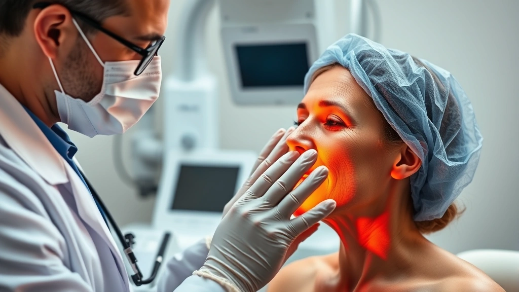
Radiation therapy stands as one of the most effective cancer treatment modalities, with millions of patients benefiting from its targeted approach to destroying malignant cells. However, the therapeutic benefits come with a spectrum of side effects that patients and healthcare providers must carefully manage. Understanding the physical manifestations of radiation therapy, particularly the visible skin changes that occur during and after treatment, is essential for informed decision-making and proper wound care management.
The skin represents the body’s largest organ and serves as the first barrier against external radiation exposure. When high-energy radiation beams penetrate the skin to reach internal tumors, they inevitably affect the surrounding healthy tissue. This article explores the comprehensive effects of radiation therapy on skin health, what medical experts reveal about these changes, and evidence-based strategies for managing and minimizing long-term complications.
Understanding Radiation-Induced Skin Changes
Radiation-induced skin damage occurs through a process called radiation dermatitis, which develops as ionizing radiation damages DNA in epidermal and dermal cells. According to research published through the National Cancer Institute, approximately 95% of patients receiving external beam radiation therapy experience some degree of skin reaction. The severity ranges from mild erythema to severe ulceration, depending on multiple factors including radiation dose, treatment duration, skin type, and individual sensitivity.
The mechanism behind these changes involves both direct cellular damage and inflammatory responses. When radiation energy deposits in skin tissue, it creates free radicals that damage cell membranes and genetic material. The body’s inflammatory response, while necessary for healing, can amplify tissue damage and lead to prolonged recovery periods. Understanding this biological process helps explain why skin changes persist long after treatment completion.
Healthcare providers use the Common Terminology Criteria for Adverse Events (CTCAE) scale to classify radiation dermatitis severity. Grade 1 represents faint erythema, Grade 2 involves moderate erythema with possible edema, Grade 3 includes severe erythema with blistering and weeping, and Grade 4 encompasses tissue necrosis and severe ulceration. This standardized classification enables consistent documentation and comparison of treatment outcomes across medical institutions.
Different body regions exhibit varying susceptibility to radiation damage. Areas with naturally moist skin, such as skin folds, the perineum, and areas under the breast, experience more severe reactions due to increased moisture retention and friction. Additionally, patients with darker skin tones may experience delayed recognition of radiation dermatitis, as erythema appears less visible on pigmented skin, potentially leading to underreported severity.
Acute Radiation Dermatitis: The Immediate Response
Acute radiation dermatitis typically manifests within the first two weeks of treatment initiation, with most patients experiencing visible changes by the third week of therapy. The progression follows a predictable pattern that medical professionals can anticipate and prepare patients to manage effectively.
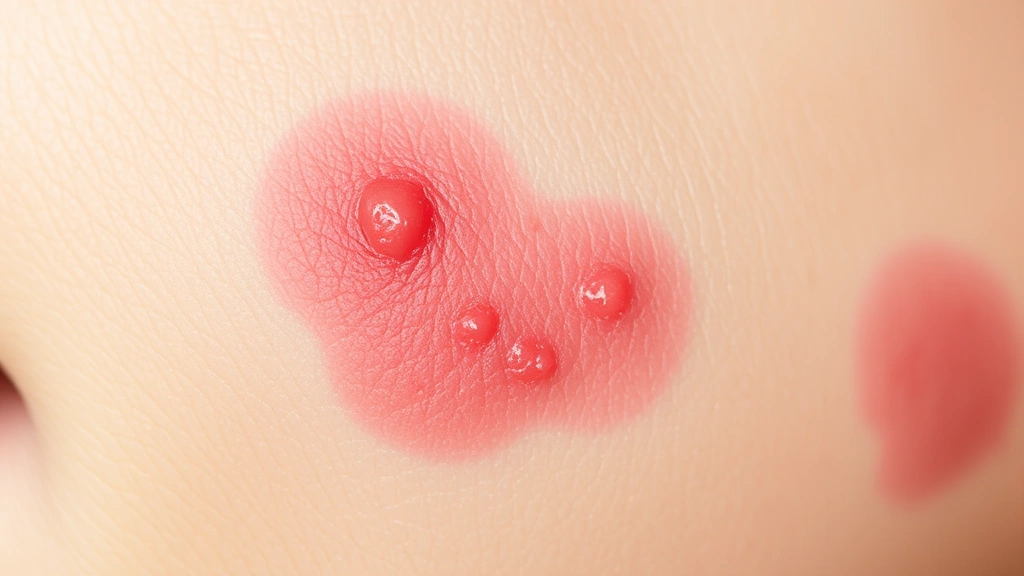
Early-phase reactions begin with transient erythema, often described by patients as a sunburn-like appearance. This initial redness results from increased blood flow to the irradiated area as the body attempts to repair damaged tissue. Patients frequently report mild itching and warmth at this stage. Some individuals experience temporary hair loss (epilation) in the treated area, which occurs because radiation damages rapidly dividing hair follicle cells.
As treatment progresses into weeks two and three, peak reactions develop. Patients may notice increasing erythema intensity, dry desquamation (dry, flaking skin), and localized edema. Some experience pruritus severe enough to interfere with sleep and daily activities. At this stage, proper skin care becomes crucial to prevent secondary infections and minimize discomfort. Healthcare providers recommend gentle cleansing with lukewarm water and fragrance-free products, avoiding tight clothing over treatment areas, and using prescribed topical medications.
In cases of severe acute dermatitis, moist desquamation develops, characterized by weeping, blistered skin and potential open wound formation. This complication occurs in approximately 10-15% of patients receiving standard radiation doses, though rates increase significantly for patients receiving higher doses or receiving concurrent chemotherapy. Moist desquamation requires intensive wound management, including sterile dressings, careful monitoring for infection, and sometimes temporary treatment interruption to allow healing.
Recovery from acute dermatitis typically occurs within 2-4 weeks after treatment completion, though individual timelines vary considerably. Factors influencing recovery speed include age, overall health status, nutritional status, and concurrent medications. Patients with diabetes, vascular disease, or compromised immune systems often experience prolonged healing periods.
Late Effects and Chronic Skin Damage
While acute dermatitis resolves within weeks to months, late radiation effects can develop months or even years after treatment completion. These chronic changes reflect permanent alterations in skin structure and function that require long-term management strategies. Medical experts emphasize that late effects represent an important consideration in radiation therapy planning, particularly for patients with long life expectancies.
The primary late effects include telangiectasia, the appearance of dilated blood vessels visible as red or purple lines on the skin surface. This occurs because radiation damages the endothelial cells lining small blood vessels, causing them to become fragile and permanently dilated. Telangiectasia typically becomes apparent 6-12 months after treatment and may progress over several years. While primarily cosmetic, these vascular changes can occasionally bleed if traumatized.
Fibrosis represents another significant late effect, characterized by excessive collagen deposition and tissue hardening in the radiation field. As fibroblasts respond to chronic inflammation, they produce excessive collagen that becomes poorly organized, creating a firm, sometimes painful area of thickened skin and underlying tissue. Fibrosis can restrict movement if it occurs near joints and may cause functional impairment beyond cosmetic concerns.
Pigmentation changes occur in approximately 40-50% of patients receiving radiation therapy. Hyperpigmentation (darkening) is more common than hypopigmentation (lightening), particularly in individuals with darker baseline skin tones. These changes reflect altered melanin production by surviving melanocytes and usually stabilize within 12-24 months after treatment completion. Some patients experience permanent pigmentation changes that require cosmetic management.
Research from peer-reviewed radiation oncology literature indicates that late skin toxicity correlates with total radiation dose, dose per fraction, and individual radiosensitivity. Doses exceeding 60 Gray (Gy) carry significantly higher risk for severe late effects. Modern treatment planning techniques attempt to minimize dose to surrounding normal tissues, thereby reducing late toxicity incidence.
A particularly concerning late effect involves radiation-induced malignancy, where cells damaged by radiation therapy develop into secondary cancers years or decades after initial treatment. While rare, this risk represents a sobering long-term consideration. Dermatologists monitor radiation therapy sites closely for any concerning skin changes that might indicate malignant transformation.
Clinical Management and Prevention Strategies
Preventing and managing radiation dermatitis requires a multifaceted approach combining preventive measures, supportive care, and targeted interventions. Medical centers have developed evidence-based protocols that significantly reduce dermatitis severity and improve patient outcomes.
Skin care protocols form the foundation of dermatitis management. Patients should cleanse the treatment area gently with lukewarm water and mild, unscented soap, patting skin dry rather than rubbing. Avoiding irritants such as perfumes, lotions containing alcohol, deodorants, and tight clothing over treatment areas helps minimize additional skin trauma. Many radiation oncology centers provide detailed written instructions and demonstrate proper skin care techniques during treatment planning.
Topical interventions have demonstrated varying efficacy in clinical trials. Hydrocortisone cream reduces inflammation during acute dermatitis phases, while silver sulfadiazine provides antimicrobial protection for areas with moist desquamation. Some centers use barrier creams containing zinc oxide or dimethicone to protect skin before treatment begins. However, applying these products before radiation may interfere with treatment delivery, so timing must be carefully coordinated with radiation oncology staff.
Emerging evidence supports the use of amifostine, a radioprotective agent that reduces free radical formation when administered before radiation therapy. However, cost and side effects limit widespread use. Similarly, pentoxifylline and vitamin E show promise in treating established fibrosis by reducing inflammatory cytokine production, though clinical benefit remains modest.
For patients experiencing severe acute dermatitis with moist desquamation, advanced wound care products including hydrogel dressings, foam dressings, and alginate products maintain appropriate moisture levels while protecting against infection. Some centers employ hyperbaric oxygen therapy for severe late fibrosis and non-healing wounds, though evidence for efficacy remains mixed.
Nutritional support plays an underrecognized role in managing radiation effects. Adequate protein intake supports tissue repair, while antioxidant-rich foods containing vitamins C and E may reduce free radical damage. Some patients benefit from nutritional counseling integrated into their cancer care team’s approach.
Rehabilitation and Recovery Support
Beyond acute treatment phase management, comprehensive rehabilitation addresses functional impairment and quality-of-life concerns resulting from radiation therapy effects. This integrative approach recognizes that skin damage often accompanies deeper tissue injury requiring coordinated rehabilitation services.
Patients experiencing fibrosis-related movement restriction may benefit from physical therapy treatment approaches that address tissue mobility and functional recovery. Physical therapists trained in cancer rehabilitation employ gentle stretching, scar tissue mobilization, and progressive strengthening to restore function and reduce pain. Physical therapy for kids receiving radiation therapy requires specialized approaches addressing developmental considerations and age-appropriate coping strategies.
For pediatric patients, pediatric physical therapy specialists integrate rehabilitation with normal developmental activities, ensuring children maintain age-appropriate function despite treatment effects. Young patients often experience better long-term outcomes when rehabilitation begins early and incorporates play-based activities that maintain motivation.
Psychological support addresses the emotional impact of visible skin changes. Many patients experience body image concerns, social anxiety, and depression related to radiation dermatitis and late effects. Mental health professionals trained in cancer survivorship help patients develop coping strategies and process feelings about treatment consequences. Support groups connecting patients with shared experiences provide valuable peer support.
Cosmetic rehabilitation offers practical solutions for patients distressed by permanent skin changes. Dermatologists specializing in radiation effects employ techniques including laser therapy for telangiectasia, topical depigmenting agents for hyperpigmentation, and cosmetic tattooing to camouflage pigmentation irregularities. While not curative, these approaches significantly improve appearance and boost patient confidence.
Long-term follow-up surveillance remains essential for detecting late complications. Dermatologists monitor radiation therapy sites annually, watching for signs of malignant transformation, progressive fibrosis, or vascular complications. Patients should report any concerning changes immediately, including new ulcerations, bleeding, or rapidly enlarging areas.
Patient Testimonials and Real-World Outcomes
Understanding radiation therapy effects requires hearing from patients who have navigated treatment and recovery. Real-world experiences illuminate the practical challenges and successes that clinical literature sometimes misses.
Many patients report that anticipatory anxiety about skin changes exceeds the actual experience. When healthcare providers thoroughly explain expected changes and provide robust support systems, patients demonstrate greater psychological resilience. One common observation involves the importance of early education: patients who understand that erythema and dry desquamation represent normal, expected responses cope more effectively than those surprised by these changes.
Patients frequently emphasize the critical importance of consistent skin care throughout treatment. Those maintaining meticulous hygiene protocols and using recommended products report significantly better outcomes than those neglecting preventive measures. This underscores that patient education and engagement directly influence treatment success.
Long-term survivors often describe adaptation to permanent skin changes as occurring gradually over months and years. Initial distress about visible alterations diminishes as patients recognize that most changes stabilize and become less noticeable over time. Many report that acceptance develops through connection with other survivors and recognition that skin changes represent evidence of successful cancer treatment.
Some patients benefit enormously from supportive services including speech therapy for toddlers and other rehabilitation services when radiation therapy affects areas influencing communication or swallowing. Occupational therapy for kids helps pediatric patients maintain independence in self-care and daily activities despite radiation effects. These specialized services often receive insufficient attention in cancer treatment planning but substantially improve quality of life.
Importantly, patient experiences vary tremendously based on cancer type, radiation dose, treatment location, and individual factors. Some patients experience minimal skin changes while others develop severe dermatitis requiring intensive management. This variability underscores the importance of individualized treatment planning and supportive care.
FAQ
What does skin look like immediately after radiation therapy?
Immediately after treatment, skin typically appears normal or shows mild erythema resembling a light sunburn. Most visible changes develop gradually over the first 2-3 weeks of treatment as inflammation increases. Acute changes peak around the third to fourth week, with gradual improvement beginning after treatment completion.
How long does radiation dermatitis take to heal?
Acute radiation dermatitis typically resolves within 2-4 weeks after treatment completion for most patients. However, some individuals experience prolonged healing, particularly those with moist desquamation or compromised healing capacity. Complete resolution of all acute symptoms may require 6-8 weeks in severe cases.
Are radiation skin changes permanent?
Acute dermatitis resolves completely in most patients. However, late effects including telangiectasia, fibrosis, and pigmentation changes often become permanent. These late effects usually stabilize within 12-24 months after treatment, though some progression may occur over several years.
Can radiation therapy cause skin cancer?
While radiation-induced secondary malignancies are rare, they represent a documented risk, particularly when high doses are delivered to large skin areas. The incidence increases with longer follow-up time and higher radiation doses. This risk must be weighed against the benefits of treating the primary cancer.
What is the best way to care for skin during radiation therapy?
Optimal skin care involves gentle cleansing with lukewarm water and mild soap, patting skin dry, avoiding irritants and tight clothing, using recommended protective creams as directed, and maintaining adequate nutrition and hydration. Following healthcare provider instructions specifically is essential, as some interventions must be timed relative to treatment delivery.
Does everyone experience severe dermatitis?
No, dermatitis severity varies considerably among patients. Factors including radiation dose, treatment duration, skin type, age, and individual radiosensitivity influence severity. Approximately 5% of patients experience minimal reactions while others develop severe dermatitis requiring intensive management.
Can late radiation effects be treated?
Some late effects respond to treatment. Telangiectasia may improve with laser therapy, hyperpigmentation sometimes responds to depigmenting agents or laser treatment, and fibrosis may benefit from pentoxifylline, vitamin E, or hyperbaric oxygen therapy. However, reversal of all changes is not possible, and treatment focuses on symptom management and appearance improvement.