Physical Therapy for Patellofemoral Pain Relief: Evidence-Based Strategies
Patellofemoral pain syndrome (PFPS), commonly known as runner’s knee, affects millions of individuals worldwide, ranging from athletes to office workers. This condition involves pain around the kneecap (patella) and typically worsens with activities like running, jumping, squatting, or climbing stairs. The pain stems from improper tracking of the patella within the femoral groove, often caused by muscle imbalances, biomechanical dysfunction, or overuse. Understanding the underlying mechanisms and implementing targeted patellofemoral syndrome physical therapy interventions can significantly reduce pain and restore functional movement patterns.
Physical therapy has emerged as the gold standard treatment approach for patellofemoral pain relief, with research consistently demonstrating superior outcomes compared to passive interventions alone. A comprehensive rehabilitation program addresses the root causes rather than merely masking symptoms, focusing on strengthening weak muscle groups, improving flexibility, correcting movement patterns, and gradually returning to normal activities. This evidence-based approach empowers patients to take an active role in their recovery while building resilience against future injuries.
Understanding Patellofemoral Pain Syndrome
Patellofemoral pain syndrome represents a multifactorial condition with diverse etiological factors. The patella functions as a crucial component of the knee extensor mechanism, distributing forces across a large surface area to reduce stress concentration. When biomechanical alignment becomes compromised, abnormal contact pressures develop between the patella and femur, triggering inflammation and pain. Common contributing factors include hip weakness, quadriceps imbalance, excessive foot pronation, tight hip flexors, and poor running or movement mechanics.
The condition affects approximately 7-10% of the general population, with higher prevalence among runners and athletes engaging in repetitive jumping activities. Research from the National Center for Biotechnology Information indicates that women experience PFPS more frequently than men, likely due to differences in hip biomechanics and muscle activation patterns. Understanding these epidemiological patterns helps clinicians identify at-risk populations and implement preventive strategies alongside therapeutic interventions.
Pain presentation varies considerably among individuals, ranging from mild discomfort during specific activities to chronic pain affecting daily function. Some patients report anterior knee pain, while others experience pain along the medial or lateral patellar facets. This variability necessitates personalized assessment and treatment planning rather than standardized protocols, ensuring each patient receives interventions specifically targeting their unique dysfunction patterns.
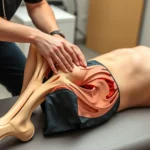
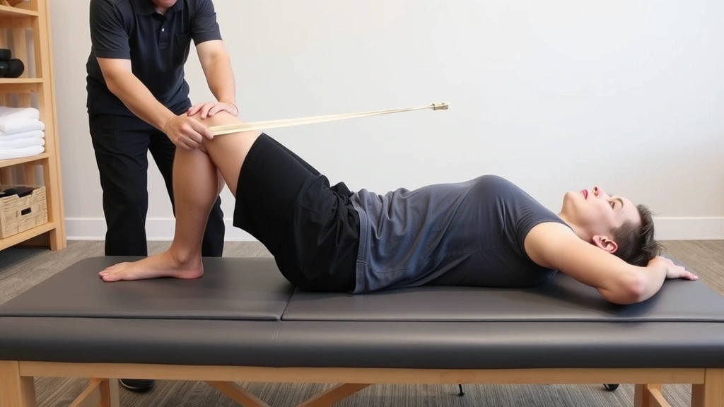
Biomechanical Assessment and Diagnosis
Comprehensive physical therapy evaluation begins with detailed history-taking and systematic biomechanical assessment. Therapists examine static posture, dynamic movement patterns, muscle strength and flexibility, and joint mobility across multiple planes. Special attention focuses on hip abductor and external rotator strength, as weakness in these muscles commonly contributes to excessive dynamic knee valgus during functional activities.
Clinical tests including single-leg squats, step-downs, and running analysis reveal compensatory movement patterns that overload patellofemoral structures. The physical therapy treatment approaches documented in modern rehabilitation emphasize movement quality assessment alongside strength testing. Imaging studies like X-rays or MRI may be warranted when red flags suggest alternative diagnoses, though many cases of PFPS can be effectively managed through clinical assessment alone.
Therapists evaluate foot biomechanics carefully, as excessive pronation can propagate forces up the kinetic chain, altering knee alignment and patellar tracking. Additionally, assessment includes hip internal and external rotation range of motion, as restrictions in these movements force compensatory stress onto the knee. Understanding the complete kinetic chain from foot through hip enables clinicians to identify all contributing factors and develop truly comprehensive treatment strategies.
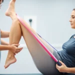
Core Strengthening Exercises
Core stability forms the foundation of lower extremity control and efficient movement patterns. The core musculature includes not only abdominal muscles but also the deep stabilizers of the lumbar spine, including the transverse abdominis, multifidus, and diaphragm. A strong, properly functioning core prevents excessive lumbar compensation and enables optimal hip positioning during dynamic activities, directly impacting patellofemoral loading patterns.
Effective core exercises progress from static holds to dynamic, multi-planar movements. Initial interventions might include prone planks, side planks, and dead bugs performed with proper breathing and muscle activation patterns. As strength improves, therapists introduce more challenging variations such as bird dogs, pallof presses, and rotational movements that mirror demands encountered during functional activities. Research from the American Physical Therapy Association emphasizes that core strength directly correlates with lower extremity injury prevention and rehabilitation success.
Progression should emphasize quality over quantity, with therapists ensuring proper muscle activation and movement control throughout all repetitions. Patients often benefit from visual feedback through mirrors or video analysis, helping them recognize and correct compensatory patterns. Home exercise programs targeting core stability should be performed consistently, ideally 4-6 days weekly, to establish neuromuscular adaptations supporting improved lower extremity mechanics.
Lower Extremity Rehabilitation
Hip strengthening represents perhaps the most critical component of patellofemoral pain rehabilitation. The gluteus medius and maximus muscles function as primary hip abductors and external rotators, controlling femoral internal rotation and adduction during single-leg stance and dynamic activities. Weakness in these muscles allows the femur to internally rotate excessively, causing dynamic knee valgus and abnormal patellar tracking.
Targeted hip strengthening exercises include side-lying hip abduction, clamshells, monster walks with resistance bands, and single-leg deadlifts. These exercises should progress from bilateral to unilateral movements, from static to dynamic variations, and from open-chain to closed-chain activities. Resistance band exercises prove particularly effective, as they provide accommodating resistance that increases as muscles move through stronger ranges of motion, optimizing strength development.
Quadriceps strengthening also requires attention, though the approach differs from traditional strength training. Rather than focusing solely on quadriceps bulk, rehabilitation emphasizes VMO (vastus medialis obliquus) activation and balanced quadriceps development. Step-ups, leg press variations, and terminal knee extension exercises with resistance bands effectively target VMO activation, promoting medial patellar stability. Avoiding excessive valgus stress during these exercises remains critical, as improper movement patterns can perpetuate the original problem.
Hamstring strengthening contributes to overall knee stability by providing dynamic posterior support and reducing reliance on quadriceps dominance. Bridging variations, prone hamstring curls, and Nordic hamstring exercises develop eccentric hamstring strength, which proves particularly important for athletes engaging in deceleration activities. Balanced quadriceps-to-hamstring strength ratios, ideally approaching 60-70% of quad strength, support optimal knee mechanics.
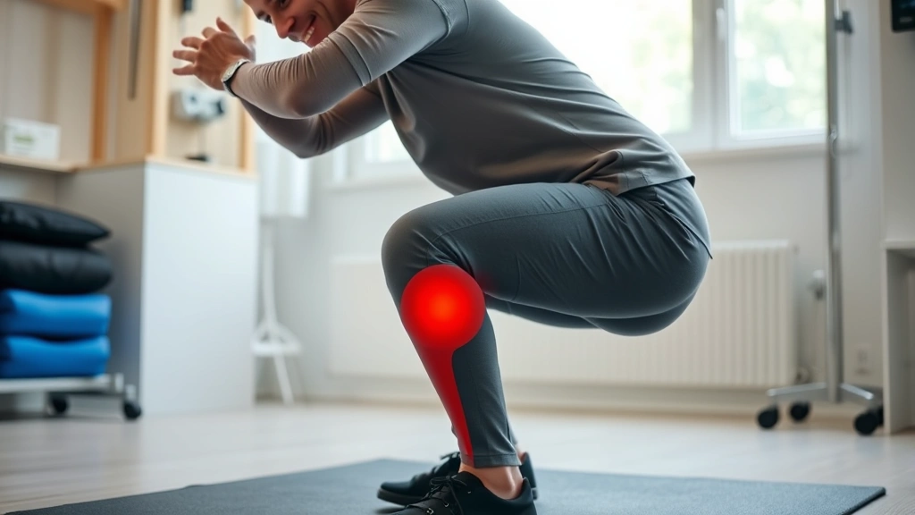
Movement Pattern Correction
Movement pattern dysfunction perpetuates patellofemoral pain and increases re-injury risk if not adequately addressed. Many patients with PFPS demonstrate excessive dynamic knee valgus during squatting, stair descent, and running—a pattern involving inward knee collapse that increases patellofemoral contact stress. Correcting this pattern requires conscious awareness, proper cueing, and repetitive practice until improved mechanics become automatic.
Therapists employ various cueing strategies to promote optimal movement patterns. Visual feedback through mirrors or video analysis helps patients understand their compensations and recognize improvements. External cueing (focusing attention on environmental targets) often proves more effective than internal cueing (focusing on body position), as it promotes more automatic, efficient movement patterns. For example, cueing patients to “push knees out” during squats encourages hip abduction and external rotation, reducing dynamic valgus.
Graduated functional training progressively introduces demands matching the patient’s return-to-activity goals. Runners benefit from running-specific training beginning with walk-to-run progressions on flat, compliant surfaces before advancing to varied terrain or speed work. Athletes engaging in cutting and jumping sports require sport-specific training emphasizing deceleration control and multi-directional movement competence. This functional progression ensures that strength and movement pattern improvements translate to meaningful activity participation.
Flexibility and Mobility Training
Muscular tightness and restricted mobility contribute significantly to patellofemoral pain by altering biomechanical alignment and limiting movement options. Hip flexor tightness, commonly occurring in sedentary individuals or those engaging in repetitive hip flexion activities, restricts hip extension and promotes anterior pelvic tilt, altering quadriceps mechanics and knee alignment. Hamstring tightness similarly restricts hip flexion, forcing compensatory lumbar spine motion and altering knee kinematics during functional activities.
Systematic flexibility training should address all major lower extremity and hip musculature. Gastrocnemius and soleus tightness contributes to excessive foot pronation, necessitating calf stretching protocols. Hip internal rotator tightness, often present in athletes engaging in repetitive external rotation activities, should be addressed through targeted stretching. Dynamic stretching before activities and static stretching after activities optimize flexibility development while minimizing injury risk.
Foam rolling and soft tissue mobilization techniques complement traditional stretching, addressing fascial restrictions and myofascial trigger points that limit movement quality. Research from ScienceDirect demonstrates that foam rolling improves range of motion and may enhance recovery between training sessions. However, these modalities should supplement rather than replace active stretching and movement-based interventions, as active flexibility development proves more functional for movement improvement.
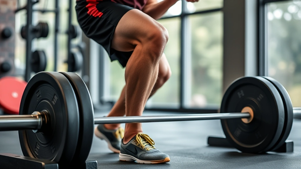
Progressive Return to Activity
Returning to sport or demanding activities represents a critical phase where rehabilitation gains must translate to functional performance. Premature return risks re-injury and perpetuation of pain, while overly conservative progression unnecessarily delays functional recovery. Evidence-based return-to-activity protocols balance these considerations through objective criteria and gradual loading progression.
Effective return-to-activity protocols typically include criteria addressing pain levels, strength symmetry, movement pattern quality, and functional task performance. Many rehabilitation specialists employ a 90% strength symmetry threshold, ensuring affected and unaffected limbs demonstrate comparable strength. Additionally, movement pattern assessment during sport-specific tasks ensures patients demonstrate proper mechanics under functional demands, not merely during controlled clinical testing.
Gradual volume progression following return-to-activity prevents re-injury while allowing tissue adaptation. The “10% rule”—increasing training volume by no more than 10% weekly—provides a conservative framework, though individual tolerance varies considerably. Runners might follow walk-to-run progressions, increasing running intervals gradually over 4-6 weeks. Athletes in cutting sports progress from straight-line movements to multi-directional activities, then to sport-specific demands at increasing intensities.
Adjunctive Modalities and Treatment
While exercise and movement pattern correction form the foundation of PFPS rehabilitation, adjunctive modalities may facilitate recovery and symptom management. Patellar taping, using athletic tape to provide mechanical support and sensory feedback, can reduce pain during early rehabilitation phases, though effects diminish as strength improves. Proper taping application by trained professionals ensures optimal benefit without compromising circulation or skin integrity.
Manual therapy techniques including soft tissue mobilization, joint mobilization, and myofascial release may enhance flexibility and reduce pain, facilitating more effective exercise participation. However, manual therapy should complement rather than replace active interventions, as research demonstrates superior long-term outcomes when combined with exercise. Some patients benefit from red light therapy and photobiomodulation approaches, though evidence remains mixed regarding isolated efficacy.
Modalities like ice application immediately following activity can manage inflammation, while heat application before exercise may enhance flexibility. Nonsteroidal anti-inflammatory drugs provide temporary pain relief but should not delay rehabilitation initiation. Corticosteroid injections occasionally benefit patients with significant inflammation, though research suggests they may impair long-term outcomes if used as substitutes for rehabilitation. The American Academy of Orthopaedic Surgeons emphasizes that conservative management through physical therapy should precede more invasive interventions.
Some therapists incorporate neuromuscular electrical stimulation (NMES) to facilitate quadriceps activation, particularly in patients demonstrating significant quadriceps inhibition. However, NMES should complement rather than replace voluntary exercise, as voluntary muscle activation produces superior strength development and motor control improvements. Dry needling and acupuncture may provide temporary symptom relief in some patients, though mechanisms remain incompletely understood.
FAQ
How long does patellofemoral pain rehabilitation typically require?
Recovery timelines vary considerably depending on pain severity, chronicity, compliance with rehabilitation, and individual healing capacity. Acute cases may improve within 4-6 weeks of consistent physical therapy, while chronic presentations may require 8-12 weeks or longer. The key involves progressive loading of tissues, gradual return to functional activities, and sustained adherence to home exercise programs. Most patients experience significant improvement within 6-8 weeks of evidence-based rehabilitation, though complete resolution may require 3-6 months.
Can patellofemoral pain syndrome be prevented?
Yes, numerous preventive strategies effectively reduce PFPS risk. Maintaining hip and core strength through regular exercise, ensuring proper running mechanics, gradually progressing training volume, and wearing appropriate footwear all contribute to injury prevention. Athletes should incorporate periodized training programs including adequate recovery, cross-training, and flexibility maintenance. Those with risk factors like hip weakness or foot biomechanical issues may benefit from preventive physical therapy assessment and targeted strengthening.
Is surgery necessary for patellofemoral pain syndrome?
Surgery is rarely necessary for PFPS, with research indicating that 90% of patients improve through conservative management. Physical therapy should always precede surgical consideration, as exercise-based rehabilitation addresses underlying biomechanical dysfunction. Surgical options like arthroscopy or patellar realignment are reserved for rare cases failing conservative treatment, and outcomes following surgery are not superior to comprehensive rehabilitation in most instances.
Should I avoid running with patellofemoral pain?
Complete activity cessation is unnecessary and potentially counterproductive. Instead, activity modification involves reducing running volume and intensity while maintaining other forms of cardiovascular conditioning like swimming or cycling. As rehabilitation progresses, gradual return-to-running programs reintroduce running in controlled, progressive manner. The goal involves training through pain rather than training through injury—maintaining activity at tolerable levels while systematically addressing underlying dysfunction.
What role does footwear play in patellofemoral pain management?
Appropriate footwear significantly influences lower extremity biomechanics and patellofemoral loading. Individuals with excessive foot pronation may benefit from stability shoes or custom orthotics controlling pronation. Conversely, those with supinated feet may require neutral or cushioned shoes. Gait analysis by trained professionals helps identify footwear needs, though shoe selection should complement rather than replace rehabilitation addressing underlying weakness and movement dysfunction.
Can physical therapy be done at home without professional supervision?
While home exercise programs form critical components of rehabilitation, initial professional assessment and guidance prove essential. Physical therapists evaluate individual presentations, identify specific dysfunction patterns, and prescribe targeted interventions. They monitor movement quality, provide corrective feedback, and progress exercises appropriately. Once rehabilitation direction is established, home programs become increasingly important, though periodic professional reassessment ensures continued progress and appropriate progression.